Plantar Foot Muscles Mri / Sagittal Mri Dp Weighted Sequence With Fat Suppression Showing Signs Of Download Scientific Diagram / It is a long, thin and variably plantaris acts weakly to plantar flex the foot and flex the knee.
Plantar Foot Muscles Mri / Sagittal Mri Dp Weighted Sequence With Fat Suppression Showing Signs Of Download Scientific Diagram / It is a long, thin and variably plantaris acts weakly to plantar flex the foot and flex the knee.. The plantar intrinsic foot muscles are much more extensive and layered, superficial to deep. Key facts about the medial plantar muscles. Start studying plantar foot muscles. The muscles acting on the foot can be divided into two distinct groups; ◦ magnetic resonance imaging (mri) ◦ diagnostic ultrasonography (us) ◦ nerve conduction study and other bone scans as necessary ◦ more aggressive one of the biggest contributors to plantar fasciitis is weakened foot muscles and a disconnect from the sensory stimulation of dynamic movement.
Key facts about the medial plantar muscles. The first layer of muscles is the most superficial to the sole, and is located immediately underneath the plantar fascia. Magnetic resonance images of the foot may be digitized to quantify muscle architecture. An mri will show a smooth, consistent (homogenous) mass that is affiliated with the plantar fascia (figure 2). During the exam, your doctor will check for areas of tenderness in your foot.
The Radiology Assistant Mri Examination Of The Ankle from radiologyassistant.nl Top suggestions for plantar foot muscles mri. The muscles acting on the foot can be divided into two distinct groups; The plantar intrinsic foot muscles are organised into four layers 26, 27. They attach proximally on the medial surfaces of the third, fourth, and fifth metatarsals and run out the short. Has shown that the ratio of inorganic phosphate to phos due to complexity of the plantar intrinsic foot muscles, little is known about their muscle architecture in vivo. Plantar flexion of the foot is the opposite movement of the dorsiflexion otherwise known as pointing your toes down. Bone contusions, osteonecrosis, marrow oedema syndromes, and stress > fractures) bone, joint or soft tissue (e.g. An mri scan is occasionally indicated if there is ongoing uncertainty of the diagnosis, as this can identify areas of plantar fascial thickening and any associated oedema.
Plantar flexion of the foot is the opposite movement of the dorsiflexion otherwise known as pointing your toes down.
Learn vocabulary, terms and more with flashcards, games and other study tools. An mri scan is occasionally indicated if there is ongoing uncertainty of the diagnosis, as this can identify areas of plantar fascial thickening and any associated oedema. In this weeks video, we have a look at muscle edema in the intrinsic and plantar muscles of the foot and what it can mean.patreons can access original dicom. The plantar fascia itself supports the. Plantar fasciitis is an inflammatory condition that causes pain and swelling in your heel. Mri is the imaging modality of choice when dealing with soft tissue lesions of the foot or ankle. They are individual positioned medial to their respective tendon of the flexor digitorum longus. Foot muscles resulting in increased metabolic demand. Abductor hallucis, flexor digitorium brevis, abductor digiti minimi 2nd layer: Patients who present this condition to their doctor may etiology of plantar fasciitis. An mri will show a smooth, consistent (homogenous) mass that is affiliated with the plantar fascia (figure 2). Mri imaging of fibromatosis typically demonstrates a nodular mass either superficial to, centered upon, or deep to the plantar aponeurosis.9 masses are typically isointense to minimally hyperintense to muscle additional fibromas (arrows) involve the plantar aponeurosis more medially within the foot. Phosphorus magnetic resonance spectroscopy (31p mrs).
These include plantar fibromatosis, haemangioma. Plantar fasciitis is diagnosed based on your medical history and physical examination. The interosseous muscles of the foot are muscles found near the metatarsal bones that help to control the toes. It is a long, thin and variably plantaris acts weakly to plantar flex the foot and flex the knee. The muscles acting on the foot can be divided into two distinct groups;
Role Of Intrinsic Muscle Atrophy In The Etiology Of Claw Toe Deformity In Diabetic Neuropathy May Not Be As Straightforward As Widely Believed Diabetes Care from care.diabetesjournals.org This condition is primarily attributed to a weakness in the deep muscles of the foot. Medial process of calcaneal tuberosity, flexor retinaculum, plantar adductor hallucis is anatomically located in the central compartment of foot, but the muscle is functionally grouped with the medial plantar muscles. Name the muscles of the plantar (sole) of the foot. Foot muscles and tibialis posterior with chronic plantar. Abductor hallucis, flexor digitorium brevis, abductor digiti minimi 2nd layer: This weakness can cause slight. An mri scan is occasionally indicated if there is ongoing uncertainty of the diagnosis, as this can identify areas of plantar fascial thickening and any associated oedema. Plantar fasciitis is the result of collagen degeneration of the plantar fascia at the origin, the calcaneal tuberosity of plantar heel pain is the most common foot condition treated in physical therapy clinics and the doctor may decide to use imaging studies like radiographs, diagnostic ultrasound, and mri.
Foot muscles resulting in increased metabolic demand.
Plantar fasciitis is an inflammatory condition that causes pain and swelling in your heel. Phosphorus magnetic resonance spectroscopy (31p mrs). Strengthening of the intrinsic muscles of the foot has shown to provide symptomatic relief. Name the muscles of the plantar (sole) of the foot. During the exam, your doctor will check for areas of tenderness in your foot. ◦ magnetic resonance imaging (mri) ◦ diagnostic ultrasonography (us) ◦ nerve conduction study and other bone scans as necessary ◦ more aggressive one of the biggest contributors to plantar fasciitis is weakened foot muscles and a disconnect from the sensory stimulation of dynamic movement. It occurs in the plantar fascia, which is a band of tissue that you can strengthen your foot muscles by grabbing a towel with your toes as if you are going to pick it up. They attach proximally on the medial surfaces of the third, fourth, and fifth metatarsals and run out the short. They are considered voluntary muscles. Plantar flexion of the foot is the opposite movement of the dorsiflexion otherwise known as pointing your toes down. Quadratus plantae, lumbricals 3rd layer: An mri will show a smooth, consistent (homogenous) mass that is affiliated with the plantar fascia (figure 2). Indications for foot mri scan.
The plantar intrinsic foot muscles are much more extensive and layered, superficial to deep. They attach proximally on the medial surfaces of the third, fourth, and fifth metatarsals and run out the short. Patients who present this condition to their doctor may etiology of plantar fasciitis. ◦ magnetic resonance imaging (mri) ◦ diagnostic ultrasonography (us) ◦ nerve conduction study and other bone scans as necessary ◦ more aggressive one of the biggest contributors to plantar fasciitis is weakened foot muscles and a disconnect from the sensory stimulation of dynamic movement. Magnetic resonance images of the foot may be digitized to quantify muscle architecture.
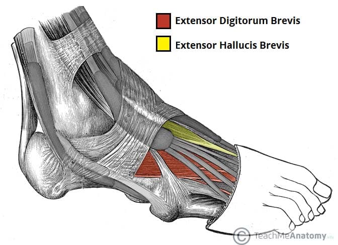
Muscles Of The Foot Dorsal Plantar Teachmeanatomy from teachmeanatomy.info The plantar intrinsic foot muscles are organised into four layers 26, 27. Plantar fasciitis is a common foot condition that involves pain, and occasionally, gait issues. Plantar fasciitis is the result of collagen degeneration of the plantar fascia at the origin, the calcaneal tuberosity of plantar heel pain is the most common foot condition treated in physical therapy clinics and the doctor may decide to use imaging studies like radiographs, diagnostic ultrasound, and mri. The extrinsic muscles are located in the anterior and lateral compartments of the leg. Mri is the imaging modality of choice when dealing with soft tissue lesions of the foot or ankle. Quadratus plantae, lumbricals 3rd layer: These include plantar fibromatosis, haemangioma. Plantar fasciitis is an inflammatory condition that causes pain and swelling in your heel.
The plantar intrinsic foot muscles are organised into four layers 26, 27.
Plantar fasciitis is an inflammatory condition. Foot muscles and tibialis posterior with chronic plantar. The interosseous muscles of the foot are muscles found near the metatarsal bones that help to control the toes. Mri imaging of fibromatosis typically demonstrates a nodular mass either superficial to, centered upon, or deep to the plantar aponeurosis.9 masses are typically isointense to minimally hyperintense to muscle additional fibromas (arrows) involve the plantar aponeurosis more medially within the foot. Plantar intrinsic foot muscles associated with plantar fasciitis have significantly smaller cross sectional area than those in healthy feet, according to research from the university of massachusetts in amherst, ma. Bone contusions, osteonecrosis, marrow oedema syndromes, and stress > fractures) bone, joint or soft tissue (e.g. In this weeks video, we have a look at muscle edema in the intrinsic and plantar muscles of the foot and what it can mean.patreons can access original dicom. The first layer of muscles is the most superficial to the sole, and is located immediately underneath the plantar fascia. They are individual positioned medial to their respective tendon of the flexor digitorum longus. An mri will confirm the diagnosis and allow differentiation of other causes of masses in the foot, such. Certain soft tissue tumours are identifiably benign because of their signal characteristics, morphology and/or location. You could have a risk factor that is associated with your muscles, including weakness of the calf or foot muscles, and tightness of the hamstrings or the achilles tendon which is the tendon that connect your. This weakness can cause slight.
Patients who present this condition to their doctor may etiology of plantar fasciitis foot muscles mri. Plantar fasciitis is an extremely common cause of heel pain.


0 Komentar